


Description
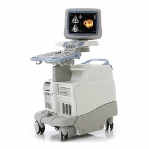
GE Vivid E95 Ultrasound Machine
The GE Vivid E95 is the premium cardiovascular ultrasound system that takes the position of the GE Vivid E9 by integrating a new and advanced software-based image processing system called cSound, a new XDclear Matrix transducer which includes volume 4D TEE options, and advanced 4D capabilities. With more flexibly programmable and powerful “cSound”, the GE Vivid E95 delivers more enhanced auto image optimization, 2D Auto EF, AFI Productivity Package, AFI Stress, and Scan Assistant Pro than those of the GE Vivid E9.
The GE Vivid E95 also achieves better ergonomics through its reduced weight, its more intuitive control panel featuring a large 12″ UHD multi-touch LCD screen, and its freely-adjustable high quality 22″ high-resolution widescreen OLED monitor. Although the GE Vivid E95 is mainly designed as a dedicated cardiovascular system, it covers a wide range of clinical applications through different types of transducers: adult cardiac, pediatric cardiac, fetal/obstetrics, abdominal (including renal, GYN/pelvic), pediatrics, small organ (including breasts, testes, and thyroid), adult and neonatal cephalic, peripheral vascular, musculoskeletal conventional, urology/prostate, transesophageal, transrectal, transvaginal, and intraoperative (including vascular, thoracic/cardiac, and abdominal).
GE Vivid E95 Dimensions and Weight:
• Width: 544 mm, 21 3/4″
• Depth: 844 mm, 33 1/4″
• Height: 1230 mm – 1670 mm, 48 3/8″ – 65 3/4″ (up/down mechanism LCD arm)
• Weight: 126 kg , 278 lbs
GE Vivid E95 Specifications:
• Infinite number of effective channels
• Minimum field-of-view range (depth): 0 – 2 cm (zoom) (probe dependent)
• Maximum field-of-view range (depth): 0 – 50 cm (probe dependent)
• Width range: 10 – 120 degrees
• Continuous dynamic receive focus/continuous dynamic receive aperture
• Continuous dynamic transmit focus
• Adjustable dynamic range, infinite upper level
GE Vivid E95 Electrical power:
• Nominal input voltage: 100-240 VAC, 50/60 Hz
• Typical power consumption: 500 W @ default cardiac preset with M5Sc
• Rated power consumption: 700 W
GE Vivid E95 Review:
The GE Vivid E95 and the Vivid E90 are GE’s new premium cardiovascular systems upgraded from the GE Vivid E9, and empowered by GE’s new cSound software beamformer and image reconstruction platform. Along with new cSound plaform, another major improvement of the GE Vivid E95 and the Vivid E90 over the Vivid E9 are 3 XDclear transducers, namely the M5Sc-D, the C2-9-D, and the C1-6-D who each add enhanced resolution, sensitivity and penetration (up to 50 cm).
Though the Matrix 4D and Matrix 4D TEE transducers seem identical to those of the Vivid E9, the newly added dedicated beamforming software on the GE Vivid E95 provides a much improved 4D cardiac capability. By combining cSound and the M5Sc XDclear transducer, the operator can expect greatly improved 2D and Doppler cardiac images.
The GE Vivid E95 is one of the few systems that integrates a large 22″ high-resolution OLED monitor. The OLED monitor offers a better clarity and resolution and is part of the next generation of monitors beyond LCD. In addition, the larger 12″ LCD touchscreen will provide the operator with a more comfortable and efficient workflow. The range of transducers offered by the GE Vivid E95 is almost similar to the Vivid E9, with the exception of a high-frequency linear transducer, which will soon be added to fully cover vascular applications.
If 4D echocardiography is a reason for choosing the GE Vivid E95, then the Philips EPIQ 7 is also a great option to consider. Though it has a smaller Matrix 4D Volume transducer, it bolsters a longer proven period of 4D echocardiography. If 4D echocardiography is not a reason for purchasing a premium cardiac system, then the GE Vivid E90 can be an alternative option. Besides the GE Vivid E95 offering a 4D option, the two systems are the same. Also, if considering the price and system reliability, GE’s factory-refurbished Vivid E9 with a 4D package can be a great option as well.
GE Vivid E95 Probes/Transducers:
Cardiac Sector Probe
M5Sc-D [1.4 – 4.6MHz]
6S-D [ 2.4 – 8.0MHz]
12S-D [ 4.0 – 12.0MHz]
4D Cardiac Sector Probe
4V-D [1.5 – 4.0MHz]
Linear Probe
9L-D [2.4 – 10.0MHz]
11L-D [4.5 – 12.0MHz]
Intraoperative Probe
L8-18i-D [5.0 – 15.0MHz]
Convex Probe
C1-6-D [1.5 – 6.0MHz]
C2-9-D [2.3 – 8.4MHz]
Micro Convex Probe
8C [4.0 – 8.0MHz]
Endo Cavity Probe
IC5-9-D [3.3 – 8.6MHz]
Non-imaging Doppler Probe
P2D [2 MHz]
P6D [6.3MHz]
Audult TEE Probe
6Tc [3.0 – 8.0MHz]
6Tc-RS [3.0 – 8.0MHz]
Pediatric TEE Probe
9T [3.0 – 10.0MHz]
9T-RS [3.0 – 10.0MHz]
Volume TEE Probe
6VT-D [3.0 – 8.0MHz]
GE Vivid E95 Features:
• High resolution 22″ wide OLED monitor
• High resolution 12″ wide LCD touch screen
• 4 active probe ports and one CW port
• ECG port
• Integrated HDD
• Multiple USB ports (front/back)
• Integrated DVD-R multi drive (optional )
• On-board storage for B/W thermal printer
• Integrated speakers for premium sound
• Integrated locking mechanism that provides rolling lock and caster swivel lock
• Integrated cable management
• Easily accessible removable air filters for cleaning
• Front and rear handles
• Side storage trays
• Rear storage trays/baskets
• Hand rest
• Automatic Optimization
• CrossXBeam
• Speckle Reduction Imaging (SRI-HD)
• Fine Angle Steer
• Coded Harmonic Imaging
• Tissue velocity M-mode
• Continuous wave Doppler
• Tissue M-mode
• Pulsed wave Doppler
• Anatomical M-mode
• Curved anatomical M-mode
• Tissue velocity imaging
• Tissue velocity Doppler
• B-flow
• Raw Data Analysis
• Real-time automatic Doppler calcs
• Tissue Tracking Mode
• Contrast Imaging LVO Contrast
GE Vivid E95 Peripheral Options:
• Sony Digital UP-D711 Termal Printer
• Sony Fixture Kit for Digital UP-D711 Thermal Printer
• Sony Digital UP-D25 Color Thermal Printer
• Sony Digital UP-D897 BW Thermal Printer
• Mitsubishi P93W/E Thermal Printer
• Mitsubishi P95DW Thermal Printer
• USB B/W video printer with control from system
• External USB printer connection
• Direct streaming DVR (Sony® HVO-550MD)
• 16 GB encrypted memory stick
• 2 TB USB hard drive (2 x 2 TB SATA II hard drives mirrored for data redundancy)
• Three-pedal configurable footswitch
• DVI-I
• Ethernet – 10 Mbps, 100 Mbps, 1 Gbps
• Multiple USB 2.0 ports
• RS-DLP Probe Adapter for TEE Probe
• Integrated gel holders
• User-configurable probe holders
• ECG AHA/IEC® Cables
GE Vivid E95 Supplies:
• Aquasonic ultrasound gel
• Sono ultrasound wipes
• Sony UPP-110HG thermal printing paper
• Sony UPC-21L color thermal printing pack
• Mitsubishi KP95HG thermal roll paper (new)
• Mitsubishi KP65HM-CE High density thermal paper
GE Vivid E95 ports:
• DVI-I
• Ethernet – 10 Mbps, 100 Mbps, 1 Gbps
• Multiple USB 2.0 ports
GE Vivid E95 image storage:
• 4D virtual store
• On-board database of patient information from past exams
• Storage formats:
– DICOM®-compressed or uncompressed, single/multi-frame, with/without raw data, storage via clipboard and/or seamlessly directly to destination device
– Transfer/ “Save As” JPEG, MPEG, AVI, DICOM, Raw DICOM and VolDicom formats
• Storage devices:
– USB memory stick: 16 GB
– CD-RW storage: 700 MB (DVD option required)
– DVD storage: -R (4.7 GB) (DVD option required)
– Hard drive image storage: 0.5 TB
• Compare old images with current exam
• Reload of archived data sets
• Activation control of USB devices
GE Vivid E95 Applications:
• Cardiac
• Stress (incl. Exercise, QStress and LVO Stress) (optional )
• Abdominal (incl. renal)
• Vascular (incl. carotid, LEA, LEV, UEA, UEV, aortoiliac)
• Fetal heart
• Pediatric
• Neonatal
• Neonatal head
• Small parts
• Thyroid
• Breast
• Musculoskeletal
• Intra Operative
• Transcranial
• Scrotal
• Urology (incl. pelvic)
• Rodent ( incl. rats and mice for research)
• Transesophageal
• OB/GYN
• Coronary
• Contrast (optional)
• Contrast low MI (optional)
• LVO contrast
GE Vivid E95 FAQs:
Auto Optimization is a one-touch image optimization function that a user uses to optimize the image, based on the actual B-mode image or Pulse wave Doppler data. The function works based on preset levels (Low, Medium, and High) and allows users to pick a preference for the contrast enhancement in the resulting image. Low does the least amount of contrast enhancement, high does the most. Auto Optimization is available in single or multi-image, on live, frozen or CINE images (in B-Mode only), while in zoom, and in Spectral Doppler. Auto Optimization in PW Doppler Mode optimizes the spectral data. Auto adjusts the Velocity Scale (live imaging only), baseline shift, dynamic range, and invert (if preset). Upon deactivation, the spectrum is still optimized.
B-Flow utilizes grayscale imaging to visualize a blood flow with different gray intensities according to the reflectors’ speed and hemodynamics. B-Flow is less dependent on the user or scanning angle, while Color Doppler is heavily dependent on the scanning angle. It also provides a higher frame rate and spatial resolution than Color Flow. B-Flow may help visualize vessel-wall irregularities, kidney perfusion, liver and spleen vasculature, and bladder reflux or jets.
Anatomical M-Mode helps improve the accuracy of arrhythmia assessments and cardiac measurements. It enables to provide M Mode images truly perpendicular to the ventricular septum.
Virtual Apex provides a wider FOV in the nearfield. Available on sector probes.
Raw Data is a software tool that enables image processing, quick data re-acquisition, and image analysis with the same resolution and same frame rates of original images. Raw Data shortens exam duration, improves clinical workflow by post-processing, and reduces a time to put the probe on a patient.
Matrix Array Volume Technology is the most advanced transducer technology in the ultrasound industry. It overcomes the limitation of volume achieving rate, improves the image quality of coronal view, and enables unlimited post-processing. The matrix array has a 2-dimensional arrangement of transducer elements like squares on a chessboard. This allows control over the sound beam in two perpendicular angles. With that, one can steer and focus the sound beam on a whole volume.
Auto IMT automates the measurement of the intima-media thickness of the vessel. Auto IMT helps keep tracking atherosclerosis diseases from the early stage as it is developed.
Tissue Velocity Imaging (TVI) is a myocardial Doppler Imaging with a color overlay on the tissue image that is available on the sector probes. Tissue color overlay can be removed to show just the 2D image, while still retaining the tissue velocity information. Tissue Velocity Imaging (TVI) is an ultrasound-based technique used for quantitative analysis of the cardiac function and has earlier been evaluated according to myocardial velocities. The technique is the same as for Doppler echocardiography, which measures flow velocities. Tissue signals, however, have higher amplitude and lower velocities, and the signals are extracted by using different filters and gain settings.
TSI (Tissue Synchronization Imaging) is a parametric imaging tool derived from Tissue Velocity Imaging (TVI), which portrays regional asynchrony on 2D echocardiographic pictures. This image is portrayed by transforming the timing of regional peak velocity during the ejection phase into color codes and overlying the moving myocardium. This allows immediate visual identification of a regional delay in systole, while a quick quantitative measurement of regional delay can be taken.
Strain and Strain Rate are for evaluation of regional myocardial function, and for assessing synchronicity and guidance during biventricular pacing procedures. Strain and Strain Rate utilizes the TVI mode. Strain calculates and color-codes the extent of tissue deformation (lengthening or shortening) relative to the original size over a given time interval, typically the systole. Strain rate calculates and color-codes the deformation per unit time i.e. the speed at which the tissue deformation occurs. Strain rate is calculated as the spatial gradient of velocity data.
Auto EF is the function that automatically measures LV ejection fraction. Along with the Automated Function Imaging (AFI) feature, Auto EF maximizes time efficiency by the reproducible quantitative output that increases the physician’s diagnostic confidence.
Automated Function Imaging (AFI) is a decision support tool for regional and global assessment of the LV systolic function. AFI is a tool derived from 2D Strain, which calculates the myocardial tissue deformation based on feature tracking on 2D greyscale loops.
Left Ventricular Contrast imaging: The LV Contrast and LVO Stress applications are optimized for endocardial border detection and assessment of wall motion and wall thickening.
Contrast Low MI: enables real-time imaging, using an MI low enough to generate little tissue signal whilst generating sufficient signal from microbubbles. This allows continuous imaging as the low MI avoids significant bubble destruction. A flash of high MI ultrasound pulses can intentionally destroy microbubbles, while, contrast replenishment is then observed to allow qualitative and quantitative assessment of the myocardium utilizing a contrast agent.
4D Auto LVQ (Automated Left Ventricular Quantification) tool enables the estimation of the left ventricular volumes and the ejection fraction in 4D data sets based on automatic border detection. The tool also enables estimation of left ventricular mass and strain (only with transthoracic 4D acquisitions). The automatic border detection is created after placing two points in an end-diastolic apical view: one at the center of the LV base and one at the apex.
4D Auto AVQ (4D Automated Aortic Valve Quantification) tool allows the user to perform computer-assisted alignment of 4D TEE images of the Left Ventricular Outflow Tract (LVOT) as well as segmentation of the LVOT and aortic root. Upon completion of the segmentation, the 4D Auto AVQ will provide metrics of the diameter and area for the aortic annulus. The 4D Auto AVQ tool is only available on Vivid E95.
4D Mitral Valve Assessment is the semi-automated MV assessment tool from Tomtec that provides the ability to include quantitative results for the mitral valve apparatus, into the patient’s exam. 4D MV-Assessment only works on stored patient data. It does not work on images that were just acquired and not yet stored.
4D Right Ventricle (RV) volume tool from Tomtec provides volume, ejection fraction, TAPSE and RV strain values from volumetric data sets. The analysis tool provides the ability to include results, from both alphanumeric values and screen captures, into the patient exam.
Quantitative analysis (Q Analysis) software package is designed for analysis of TVI related (Tissue Tracking, Strain, Strain rate, TSI) and Contrast related raw data. Q Analysis traces for velocity or derived Parameters inside defined regions of interest as a function of time.
M5Sc-D, 6S-D, 12S-D, 4V-D, 9L-D, 11L-D, L8-18i-D, C1-6-D, C2-9-D, 8C-D, iC5-9-L, P2D, P6D, 6Tc, 6Tc-RS, 6VT-D, 9T, 9T-RS
CLICK HERE TO REQUEST A PRICE QUOTE