


Description
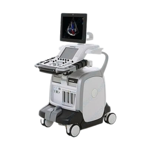
GE Vivid iQ Portable Ultrasound Machine For Sale
The GE Vivid iq is a new and upgraded GE’s Vivid line cardiovascular portable ultrasound system from the base of Vivid q and Vivid i. The Vivid iq has integrated the full touch screen control 15.6″ monitor on conventional user control panel upgrading the user’s intuitive control and operation quality. The Vivid iq applies the trackpad design instead of the trackball interface. The GE Vivid iq is light weight, and with the dedicated cart, three RS and one DLP to support 6VT-D ports are available. Optional three batteries enable to scan more than 180 min with the system battery. The GE Vivid iq enables to support cardiac, transesophageal, peripheral vascular, fetal/OB, abdominal adults, pediatric, small organ, neonatal cephalic, adult cephalic, musculoskeletal.
CLICK HERE TO REQUEST A PRICE QUOTE
GE Vivid iq Dimensions and Weight:
• Height: 64 mm (2.5″)
• Width: 390 mm, (15.35″)
• Depth: 362 mm (14.25″)
• Weight with battery: 5.2 kg (11.5 lbs)
GE Vivid iq Specifications:
• Digital beamformer with up to 974,026 effective digital channels
• Minimum field-of-view range (depth): 1 cm (probe dependent)
• Maximum field-of-view range (depth): 33 cm (probe dependent)
• Width range: 10° – 168° (probe dependent)
• Continuous dynamic receive focus/continuous dynamic receive aperture
• Adjustable dynamic range, infinite upper level
GE Vivid iq Electrical power:
• Nominal input voltage: 100-240 VAC, 50/60 Hz
• Rated power consumption: 500 VA
GE Vivid iq Probes/Transducers:
Cardiac Sector Probe
3Sc-RS [1.3 – 4.0MHz]
6S-RS [2.0 – 7.0MHz]
12S-RS [4.5 – 12.0MHz]
Linear Probe
9L-RS [2.4 – 10.0MHz]
12L-RS [4.0 – 13.0MHz]
Convex Probe
4C-RS [1.5 – 5.0MHz]
Micro Convex Probe
8C-RS [3.5 – 10.0MHz]
Endo Cavity Probe
E8Cs-RS [3.5 – 10.0MHz]
Non-imaging Doppler Probe
P2D [2 MHz]
Audult TEE Probe
6Tc-RS [3.0 – 8.0MHz]
Pediatric TEE Probe
9T-RS [4.0 – 10.0MHz]
GE Vivid iq Features:
• Tissue velocity M-mode
• Continuous wave Doppler
• Tissue M-mode
• Pulsed wave Doppler
• Anatomical M-mode
• Tissue velocity imaging
• Tissue tracking
• Tissue velocity Doppler
• Blood flow imaging
• B-flow
• Virtual convex
• Virtual apex
• Coded phase inversion
• Compound imaging
GE Vivid iq Peripheral Options:
• Sony Digital UP-D711 Termal Printer
• Sony Fixture Kit for Digital UP-D711 Thermal Printer
• Sony Digital UP-D25 Color Thermal Printer
• Sony Digital UP-D897 BW Thermal Printer
• Mitsubishi P93W/E Thermal Printer
• Mitsubishi P95DW Thermal Printer
• DVDRW
• Eight GB memory stick
• One TB USB hard drive
• USB interface (5)
• HDMI cable
• Three-pedal configurable footswitch
• Rolling bag
• ECG AHA/IEC® Cables
GE Vivid iq Supplies:
• Aquasonic ultrasound gel
• Sono ultrasound wipes
• Sony UPP-110HG thermal printing paper
• Sony UPC-21L color thermal printing pack
• Mitsubishi KP95HG thermal roll paper (new)
• Mitsubishi KP65HM-CE High density thermal paper
GE Vivid iq Console ports:
• HDMI interface
• Ethernet – 10 Mbps, 100 Mbps, 1 Gbps
• Multiple USB 2.0 ports
GE Vivid iq Cart Design:
• Three USB ports
• Six probe holders
• Four probe cable hooks
• Charge box (optional ) – to charge up to three batteries and to scan more than 180 min with four fully charged batteries
• Multi-probe box (optional ) – three RS, one DLP to support 6VT-D
GE Vivid iq image storage:
• On-board database of patient information from past exams
• Storage formats:
– DICOM®-compressed or uncompressed, single/multi-frame, with/without raw data, storage via clipboard and/or seamlessly directly to destination device
– Transfer/“Save As” JPEG, MPEG, AVI formats
• Storage devices (optional ) :
– USB memory stick: eight GB
– CD-RW storage: 700 MB (DVD option required)
– DVD storage: -RW (4.7 GB)
– Hard drive image storage: one TB
• Compare previous images with current exam
• Reload of archived data sets
GE Vivid iq Applications:
• Cardiac
• Transesophageal
• Peripheral vascular
• Fetal/OB
• Abdominal adults
• Pediatric
• Small organ
• Neonatal cephalic
• Adult cephalic
• Musculoskeletal conventional
• Musculoskeletal superficial
• Transcranial
• Transrectal
• Transvaginal
GE Vivid iq FAQs:
1. Auto Optimization is a one-touch image optimization function that a user to optimize the image based on the actual B-mode image or Pulse wave Doppler data. The function works based on preset levels (Low, Medium, and High) and allows user to pick a preference for the contrast enhancement in the resulting image. Low does the least amount of contrast enhancement, high does the most. Auto Optimization is available in single or multi image, on live, frozen or CINE images (in B-Mode only), while in zoom, and in Spectral Doppler. Auto Optimization in PW Doppler Mode optimizes the spectral data. Auto adjusts the Velocity Scale (live imaging only), baseline shift, dynamic range, and invert (if preset). Upon deactivation, the spectrum is still optimized.
2. B-Flow utilizes grayscale imaging to visualize a blood flow with different gray intensities according to the reflectors’ speed and hemodynamics. B-Flow is less dependent on the user or scanning angle, but Color Doppler heavily dependent on scanning angle, and also provides a higher frame rate and spatial resolution than Color Flow. B-Flow may help visualize vessel-wall irregularities, kidney perfusion, liver and spleen vasculature, and bladder reflux or jets.
3. Anatomical M-Mode helps improve accuracy of arrhythmia assessments and cardiac measurements. It enables to provide M Mode images truly perpendicular to the ventricular septum.
4. Virtual Apex provides wider FOV in the nearfield. Available on sector probes
5. Raw Data is a software tool that enables image processing, quick data re-acquisition, and image analysis with same resolution and same frame rates of original images. Raw Data helps shorten exam duration, improves clinical workflow by post-processing, and reduces a time to put a probe on a patient.
6. Auto IMT automates the measurement of intima-media thickness of vessel. Auto IMT helps keep tracking atherosclerosis diseases from the early stage as it is developed.
7. Tissue Velocity Imaging (TVI) is myocardial Doppler Imaging with color overlay on tissue image available on the sector probes. Tissue color overlay can be removed to show just the 2D image, still retaining the tissue velocity information. Tissue Velocity Imaging (TVI) is an ultrasound-based technique used for quantitative analysis of the cardiac function and has earlier been evaluated according to myocardial velocities. The technique is the same as for Doppler echocardiography, measuring flow velocities. Tissue signals however, have higher amplitude and lower velocities, and the signals are extracted by using different filter and gain settings.
8. Auto EF is the function that automatically measure LV ejection fraction. Along with the Automated Function Imaging (AFI) feature, Auto EF maximizes time efficiency by the reproducible quantitative output that increases physician’s diagnostic confidence.
9. Quantitative analysis (Q Analysis) software package is designed for analysis of TVI related (Tissue Tracking, Strain, Strain rate, TSI) and Contrast related raw data. Q Analysis traces for velocity or derived Parameters inside defined regions of interest as function of time
CLICK HERE TO REQUEST A PRICE QUOTECLICK HERE TO REQUEST A PRICE QUOTE