


Description
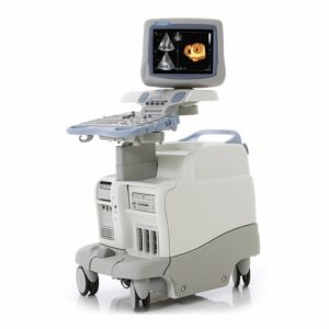
GE Voluson S8 Ultrasound Machine
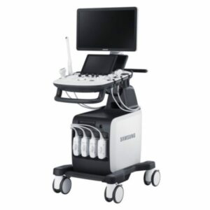
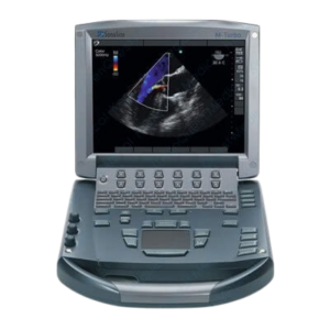
The GE ultrasound Voluson S8, is a high-end women’s health 4D ultrasound machine that has a price and features in-between the Voluson E8 and the Voluson S6. The Voluson line from GE is known worldwide for providing the best 4D imaging currently available though Samsung is catching up. The Voluson S8 and S6 look identical and even use most of the same transducers. The main difference is that the S6 uses half the channels of the Voluson S8. This produces a significant difference in image quality between the Voluson S8 and S6. The GE Voluson S8 uses the same cost-effective RS transducers as the Voluson-i portable. The Voluson S8 offers near E8 level image quality at a lower price and much smaller, lighter form factor.
All Voluson ultrasound machines are focused on women’s health, especially obstetrics, gynecology, and fertility. Secondary applications include general imaging, adult & pediatric cardiology and neonatal cardiology. Doctors looking for shared service capabilities from a GE ultrasound machine should consider the GE Logiq S8 instead.
CLICK HERE TO REQUEST A PRICE QUOTE
GE Voluson S8 Dimensions & Weight
Height: (adjustable, maximum) 1725 mm (67.9 in), (minimum) 975 mm (38.4 in)
Width: 620 mm (24.4 in)
Depth: 850 mm (33.4 in)
Weight: (no Peripherals) 90 kg (198 lbs.), approx. 325 lbs. with packaging
Voluson S8 Specifications
Digital Beamformer
335,127 system processing channel technology
Minimum Depth of Field: 0-1 cm (Zoom, probe dependent)
Maximum Depth of Field: 0-36 cm (probe dependent)
Up to 261 dB Dynamic Range adjustable by selecting 12 Dynamic Contrast Curves
GE Voluson S8 Electrical
Voltage: 100-120VAC, 220-240VAC
Frequency: 50/60 Hz ( /-2%)
Power consumption: Nominal 900VA including all options. Typical power consumption with 500VA load approx. 1.75A at 230V/50Hz without peripherals.
Thermal Output: 1200 BTU/h
GE Voluson S8 Revisions: BT11 to BT15
GE first launched the Voluson S8 in 2011. That first version was designated as BT11. “BT” is an abbreviation of “Break Through” and the number designates the year in which this version was launched. So the Voluson S8 BT11 was launched in 2011 and was in production till the next version in 2012, the Voluson S8 BT12. The Voluson S8 BT12 improved the image quality but kept the same feature set. The GE Voluson S8 BT14 added HD live (highly improved the 3D/4D image quality) as a hardware upgrade option. This option requires different hardware than the non-HD-live version. The current version of the Voluson S8 is the BT15 that adds a cellular modem kit, and MMS/Email connectivity.
All revisions of the GE Voluson S8
GE Voluson S8 (BT11)
GE Voluson S8 (BT12)
GE Voluson S8 (BT14)
GE Voluson S8 (BT15)
Popular configurations of the Voluson S8 in 2016
GE Voluson S8 (BT12) with 3 transducers
RAB4-8-RS 4D Convex
4C-RS 2D Convex
E8C-RS 2D Endovaginal
All GE Voluson S8 probes/transducers
4D Convex RAB2-5-RS [ 1 – 5 MHz ] 192 elements, 46mm, max scanning depth 30cm
4D Convex RAB2-6-RS [ 2 – 6 MHz ] 192 elements, 46mm, max depth 26cm
4D Convex RAB4-8-RS [ 2 – 8 MHz ] 192 elements, 46mm, max scanning depth 26cm
4D Endocavitary RIC5-9W-RS [ 4 – 9 MHz ] 192 elements, 11.6mm, max scanning depth 16cm
Endocavitary E8C-RS [ 4 – 10 MHz ] 128 elements, 10.7mm, max scanning depth 16cm
Convex C1-5-RS [ 2 – 5 MHz ] 192 elements, 56.1mm, max scanning depth 30cm
Convex 4C-RS [ 2 – 5 MHz ] 128 elements, 60.0mm, max scanning depth 30cm
Convex AB2-7-RS [ 2 – 8 MHz ] 192 elements, 40.0mm, max scanning depth 28cm
Micro convex 8C-RS [ 4 – 10 MHz ] 128 elements, 10.7mm, max scanning depth 16cm
Linear 12L-RS [ 4 – 12 MHz ] 192 elements, 37mm FOV, max scanning depth 8cm
Linear 9L-RS [ 3 – 8 MHz ] 192 elements, 37mm FOV, max scanning depth 14cm
Linear Matrix ML6-15-RS [ 4 – 13 MHz ] 336 elements, 50mm FOV, max scanning depth 12cm
Cardiac sector 3Sc-RS [ 1 – 4 MHz ] 64 elements, 50° FOV, max scanning depth 24cm
Pediatric cardiac sector 12S-RS [ 5 – 11 MHz ]18mm footprint, max scanning depth 12cm
Pencil Transducer P2D [ 2 MHz ] CW Split Crystal
Advanced Voluson S8 transducers: 4D and Matrix
The Voluson S8 supports a wide range of 2D and 3D probes that help medical professionals obtain great images, especially in the first trimester and in complex gynecological exams. The Voluson S8 supports four mechanical 4D transducers; 3 convex and 1 endocavitary. The advanced C1-5-RS convex offers exceptional 2D imaging. The ML6-15-RS linear probe features matrix technology for breast imaging and provides excellent spatial resolution and image uniformity in a 50 mm footprint that reduces the number of passes needed in a breast exam.
Popular GE Voluson S8 transducers
The RAB4-8-RS 4D convex gives the broadest range of penetration. The E8C-RS endocavitary and 12L-RS are both inexpensive and are popular on a wide variety of GE systems. The RIC5-9W-RS is the only 4D transducer on the Voluson S8 that offers elastography. The C1-5-RS convex is popular for 2D imaging as it offers a wider bandwidth and superior imaging.
GE Voluson S8 vs E6
The GE Voluson E6 looks nearly identical to the more expensive Voluson E8, but has half the channels. In price and image quality it is nearly identical to the Voluson S8. The S8 is a newer design and has a smaller size and weight. However the Voluson E6 can use the same transducers as the E8. These transducers offer better image quality and a wider range of transducers but at a higher price. So when choosing between the Voluson S8 and E6, it comes down to 2 choices; do you prioritize smaller size and a lower probe price? If so then the Voluson S8 is a better choice. Or if you want more transducer options and slight edge in image quality from those same advanced transducers then choose the Voluson E6. Remember to look at which revision of the E6 you are considering as there is a much larger difference between the features of the E6 over the years where as the Voluson S8 has relatively few changes from BT11 to BT15.
GE Voluson S8 Features:
These are features that are standard on the Voluson S8 BT12.
19” High-Resolution TFT LCD Monitor
Innovative user interface with onscreen menus
3 Active Probe Port
3D/4D Mode
4D Biopsy
HD-Flow
Automatic Tissue Optimization
Coded Excitation (CE)
Coded Harmonic Imaging with Pulse Inversion Technology
Tissue Doppler
Advanced SRI
CrossXBeam CRI
SonoNT
B Mode only
B Power Doppler Mode
B CFM Doppler Mode
B HD-Flow Mode
B CRI
B CRI CFM
B CRI PD
B CRI HD-Flow
B B-Flow
Focus & Frequency Composite (FFC)
Inversion mode
High-Resolution Zoom
Pan Zoom
SonoRenderStart
Steering
Tomographic Ultrasound Imaging (TUI)
Virtual Convex
VOCAL
Wide Angle on endovaginal probes
Beta-View
Patient information database
Image Archive on hard drive
3D/4D data compression (lossy/lossless)
Real-time automatic Doppler calculations
OB Measurement, Calculations & Reports
GYN Measurement, Calculations & Reports
Vascular Measurement, Calculations & Reports
Cardio Measurement, Calculations & Reports
Abdominal Measurement, Calculations & Reports
Small-Parts Measurement, Calculations & Reports
Urology Measurement, Calculations & Reports
Pediatrics Measurement, Calculations & Reports
Musculoskeletal Measurement, Calculations & Reports
Neurology Measurement, Calculations & Reports
Multigestational Calculations and Fetal Trending
CINE Memory: 140MB; up to 7,000 frames, Dual/Quad Display, Review Loop and speed
Integrated HD (500 GB)
GE Voluson S8 technology definitions:
4D Biopsy: For minimally invasive procedures like biopsies, ultrasound is a widely used method to visualize and guide the needle. 4D biopsy allows for real time control of the biopsy needle in s 3D multi-planar display. The Voluson S8 with 4D Biopsy shows the region of interest in three perpendicular planes (longitudinal, transversal and frontal sections) and can guide the biopsy needle accurately into the center of the lesion.
CE: Coded Excitation improves image resolution and penetration in the far field on the Voluson S8. This allows the user to scan at higher frequencies on technically difficult patients.
Advanced SRI: A nonlinear diffusion filtering technique that improves image quality in real time by reducing speckles. Available in all B-mode imaging on any probe compatible with the Voluson S8.
CrossXBeam CRI: This technology is borrowed from the E8. It is compound resolution imaging used to improve border and image clarity the Voluson S8.
SonoNT: Allows for semi-automatic Nuchal Translucency measurements.
FFC: Focus and Frequency Composite is a Voluson S8 technology that utilizes two different transmission frequencies and two different focal ranges in the 2D image. This function combines a low frequency to increase the penetration and higher frequency to keep the resolution high. It reduces speckles and artifacts in the 2D image to facilitate the examination of difficult-to-scan patients.
SonoRender Start: This technology on the Voluson S8 speeds up the acquisition of the fetal face in 4D.
TUI: This is a new visualization mode for 3D and 4D data sets on the Voluson S8. The data is presented as slices through the data set which are parallel to each other. An overview image, which is orthogonal to the parallel slices, shows which parts of the volume are displayed in the parallel planes. This method of visualization is consistent with the way other medical systems such as CT or MRI, present the data to the user. The distance between the different planes can be adjusted to the requirements of the given data set. In addition, it is possible to set the number of planes. The planes and the overview image can also be printed to a DICOM printer, for easier comparison
of the ultrasound data with CT and/or MRI data.
VOCAL: Imaging program opens up completely new possibilities in cancer diagnosis, therapy
planning and follow-up therapy control. It offers different functions: Manual or Semi-automatic Contour detection of structures (such as tumor lesion, cyst, prostate, etc.) and subsequent volume calculation. The accuracy of the process can be visually controlled by the examiner in a multi-planar display. A virtual shell can be specified around the contour of the lesion. The wall thickness of the shell can be defined. The shell can be imagined as a layer of tissue around the lesion, where the tumor vascularization takes place. Automatic calculation of the vascularization within the shell by 3D color histogram by comparing the number of color voxels to the number of grayscale voxels.
Beta View: allows the adjustment of the Volume O-Axis position of 3D
probes in 2D mode. The green line in the displayed symbol indicates the position of
the acoustic block. and – defines the corresponding sweep direction on the Touch
screen.
GE Voluson S8 Accessories
Sony UPD-897MD Digital Black & white thermal printer
Sony UPD-898MD Digital Black & white thermal printer
Sony UPX-898MD Digital Black & white thermal printer
Sony UPD-25MD Digital Color thermal printer
Mitsubishi P95DW Digital Black & white thermal printer
Mitsubishi CP30DW Digital Color thermal printer
Sony DVO-1000 DVD Recorder
CIVCO disposable biopsy guides (for Convex, Linear and Endo-cavity transducers)
Voluson S8 Supplies
Aquasonic ultrasound gel
Sono ultrasound wipes
Sony UPP-110HG thermal printing paper
Sony UPC-21L color thermal printing pack
Mitsubishi CK30L printing paper
Mitsubishi K95HG high gloss thermal printing paper
GE Voluson S8 ports
3 active transducer ports
3 USB Ports for External Peripherals
2 USB Ports for On-board Peripherals
Ethernet network connection
1 HDMI Out Port
1 Audio Out Port
GE Voluson S8 Standard Imaging Modes
2D-Mode
M-Mode
PW Doppler
High PRF Doppler Mode
Color Flow Doppler Mode (CFM)
Power Doppler Mode (PD)
Elastography mode
XTD-Mode
Contrast Agent Mode (Contrast)
M-Color Flow Modes (M/CF, M/HD-Flow, M/TD)
Volume Modes (3D/4D):
4D Real-Time
4D Biopsy
Real-Time Triplex
Voluson S8 Optional Imaging Modes
CW Doppler Mode (CW)
Anatomical M-Mode
B-Flow Mode (BF)
VCI-A (Advanced Volume Contrast Imaging)
VCI OmniView
STIC
3D Static
GE Voluson S8 Applications
Applications or Apps are the types of exams or studies that an ultrasound machine can do. More than this if an ultrasound machine supports a specific application it will have calculations, measurement and reporting software included to support those apps and make them useful in a clinical environment.
The GE Voluson S8 offers a broad selection of applications but is focused on OB/GYN and 4D.
Abdominal
Small Parts
Obstetrics
Gynecology
Cardiology
Urology
Vascular
Peripheral Vascular
Pediatrics
Neurology
Musculoskeletal
Breast