


Description
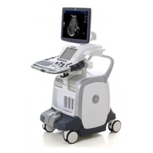
Samsung Medison HS70A
The Samsung HS70A is a high-end diagnostic ultrasound system particularly designed to fit into a general ultrasound imaging setting. The Samsung HS70A can support many applications including fetal, obstetrics, abdominal, gynecology, pediatric, small parts, neonatal cephalic, trans-rectal, trans-vaginal, MSK, urology, adult and pediatric cardiac, and peripheral vessel. The features of the HS70A allow for an in-depth OB/GYN practice, thanks to its top-tier 3D/4D technologies and automated functions that effectively reduce operational time and improve clinical workflow and productivity.
Since its merger with Samsung, the largest commercial electronics company, Samsung products have integrated Samsung’s advanced IT and semi-conductor based technologies. The result is a unique hybrid ultrasound architecture which features S-Harmonic, S-Vue transducers (CA1-7A, CV1-8A), Realistic Vue, 2D NT, 5D follicle (automatic follicle measurement), S-Detect for Breast, E-Breast and E-Thyroid utilizing static elastography, quick preset, Ez-Exam , Advanced QuickScan including color gain and color box adjustment, and TGC touch slide controls.
CLICK HERE TO REQUEST A PRICE QUOTE
Samsung HS70A Dimensions and Weight:
• Height: 1,430 ~ 1,710 mm
• Width: 557 mm
• Depth: 791 ~ 860 mm
• Weight with monitor: approx. 105.4 kg (232 lb.)
Samsung HS70A Specifications:
• Digital beamformer
• Displayed imaging depth: 0 – 38 cm (probe dependent)
• Minimum depth of field: 0 – 2 cm (probe dependent)
• Maximum depth of field: 0 – 38 cm (probe dependent)
• Continuous dynamic receive focus/continuous dynamic receive aperture
• Adjustable dynamic range from 50 to 255 dB
Samsung HS70A Electrical power:
• Voltage: 100- 240 V AC
• Frequency: 50/60 Hz
• Power Consumption
– 714.7 VA with Peripherals
– 564.7 VA without Peripherals
• Nominal Current:
– 110V: 8.2A
– 220V: 4.1A
Samsung HS70A Review:
If the end-user specializes in General Imaging, Obstetrics, Gynecology, enjoys using digital tools, and is an early adapter of new technologies, then the Samsung HS70A is a perfect choice. As a top technology leader in 3D & 4D imaging, Samsung has integrated advanced 3D/4D technology from its OB/GYN flagship system: the Samsung WS80A. Along with GE’s HDLive, Samsung’s Realistic Vue is now very popular. Also, its 2D NT and 5D Follicle features can greatly help OB/GYN medical professionals perform comfortable and accurate clinical exams. The interface, presets, and measurement packages of the HS70A easily meet market standard requirements for both OB/GYN and radiology, which will allow the end-user to complete all required exams without any difficulty.
Convex Probe
CA1-7AD
CA2-8A
CA2-9AD
CA3-10A
CF4-9
Linear Probe
LA3-12A
LA3-16A
LA2-9A
LA4-18BD
LA3-16AI
Phased Array Probe
PE2-4
PA3-8B
PA4-12B
Endocavity Probe
E3-12A
VR5-9
EA2-11B
4D Volume Probe
CV1-8AD
V5-9
LV3-14A
Ped off Probe
DP2B
CW4.0
CW6.0
DP8B
Samsung HS70A Features:
• 23″ LCD monitor (LED backlight unit)
• 10.1” LED touch screen
• Probe Active 3Ports (optional 4 Probe Port)
• Height/Rotate adjustable control panel
• Height/Tilt/Rotate the adjustable monitor
• On-board storage for peripherals
• 6 probe holders
• 4 Swivel/Lock wheels (wheel diameter: 125 mm)
• Front and rear handles
• Rear storage cover for peripheral connectors and cables
• Integrated cable management
• Integrated high-fidelity stereo speakers
• Prevention of noise of the system
• Easily removable air filter
• Integrated SSD 512GB
• Integrated DVD Multi Recordable Driver
• Full Spectrum Imaging (3 bands)
• S-Harmonic Mode(Pulse Inversion Harmonic, Coded Harmonic Imaging)
• S-Flow
• Trapezoidal Imaging
• Quick Scan™ (Automatic Optimization)
• ClearVision
• MultiVision
• Raw Data Analysis
• Post-image optimization
• Patient Information Database
• Image Archive integrated on CD/DVD and HDD drive
• Support for external USB 2.0 HDD drive
• Cine for 12,700(Max Condition) frames and Loop Review for 8,192 lines
• Auto Calc (Real-Time Automatic Doppler Calculation)
• Doppler Auto Trace
• User Configurable Measurement Menu
• Customizable Measurement Menu
• Customizable Body Maker
• Customizable User Keys
• Post-Measurement
• On-board electronic documentation
• Fully Digital Real-Time Recording
• Screen Keyboard
Samsung HS70A Peripheral Options:
• Digital BW: Mitsubishi P-95DE, Sony UP-D897, UP-X898MD, UP-D898MD
• Digital Color: Mitsubishi CP-30DW, Sony UPD25-MD
• USB BW Line: Samsung ML-2955DW
• USB Color Line: Samsung CLP-615ND
• USB Footswitch
• USB Digital ECG Kits (AHA/IEC)
• USB HDD
• USB Flash Memory Media
• USB Printer
• DVD Recorder
Samsung HS70A Supplies:
• Aquasonic ultrasound gel
• Sono ultrasound wipes
• Console Protective Cover
• Sony UPP-110HG thermal printing paper
• Sony UPC-21L color thermal printing pack
• Mitsubishi KP95HG thermal roll paper (new)
• Mitsubishi KP65HM-CE High-density thermal paper
• External USB printer connection
Samsung HS70A ports:
• Audio port (I/O)
• LAN (I/O) : Ethernet, 10/100 BASE-T
• B/W Printer port (USB type) (O)
• Patient Monitor
• VHS
• SVHS
• d-Sub (1280 x 1024)
• DVI-D (1920 x 1080)
• DVI-I (1280 x 1024)
• Patient Monitor Power (O)
• DVI port (O) for digital signals to the monitor
• ECG AUX input (optional)
• Footswitch port (USB type)
• USB port (I/O, USB 2.0) : 8 ports(front2 , rear4, USB Printer 2 )
• Microphone
Samsung HS70A image storage and documentation:
• ADVRTM
• On-board printing device control
• Selective printing on two connected printers
• SonoView II (Image Filing Package)
• Export Media: CD/DVD R/-R/RW, USB Flash, External USB HDD
• Export Format: JPEG,BMP,TIFF,DICOM
• Print Function
• Patient list and data search
• Report save available
• Post image processing available
• Caliper measurement available
• DICOM 3.0
Service: Storage/Printer/Worklist/PPS/SC
DICOM SR Structured Report
Samsung HS70A Applications:
• General
• Abdominal
• Gynecology
• Obstetrical
• OB Early
• Renal
• Fetal Heart
• Urology
• Vascular
• Small Parts
• Muskuloskeletal
• Breast
• Pediatric
• Cardiac
• TCD
Samsung HS70A FAQs:
• ClearVision
The noise reduction filter improves edge enhancement and creates sharper 2D images in order to deliver an optimal diagnostic performance. The integration of a technologically advanced Samsung product brings about a notable improvement in the quality of images. In addition, ClearVision provides application – specified optimization and advanced temporal resolution in live scan mode.
• S-Harmonic
The ordinary harmonic technology is able to provide greater image clarity from near to midfield, while also reducing signal noise. Combined with S-Vue transducers and S-Vision imaging engine, the S-Harmonic overcomes the technology limitation of the ordinary Harmonic technology, and improves the quality of the image from near to far field.
• ElastoScan
This is a diagnostic ultrasound technique that is able to display elasticity by converting the stiffness into color images, while detecting the presence of solid masses in tissues.
• Auto IMT
Auto IMT is a screening tool that is able to analyze a patient’s potential risk of cardiovascular disease. It allows easy measurements for intima-media thickness of both the anterior and posterior wall of the common carotid by one click. This simple procedure enhances exam productivity and adds diagnostic value.
• S-Vue transducers (CA1-7AD, CA2-9AD, PA1-5A, CV1-8AD)
Retaining an innovative single crystal design, S-Vue™ transducers provide more efficient piezoelectric properties. This results in wider bandwidths that enable better penetration and higher quality resolution on even challenging patients.
• S-Detect™
Upon clicking a suspicious lesion, the S-DetectTM draws the lesion borders, suggests the characteristics of the lesion and gives a hint whether the lesion is benign or malignant. S-Detect™ uses the Breast Imaging-Reporting and Data System (BI-RADS®) scores for standardized reporting and classification of lesions.
• E-Breast™
The E-BreastTM is one of ElastoScanTM’s method for displaying a color image that is superimposed on the B-mode image, which represents the elasticity of the tissue. E-Breast™ technology calculates the strain ratio between the selected target and the surrounding fatty tissue. Unlike conventional ultrasound elastography, E-Breast™ requires only one ROI to be selected by the user.
• E-Thyroid™
The ElastoScanTM displays a color image superimposed on the B-mode image, which represents the elasticity of the tissue. The E-ThyroidTM uses the pulsations of the adjacent common carotid artery (CCA), which eliminates the need for manual transducer compression while offering greater consistency in the ElastoScan™ image. E-Thyroid™ provides an elasticity contrast index that is calculated by comparing the elasticity of the lesion and the normal tissue within the ROI.
• E-Cervix™
The ElastoScanTM displays a color image superimposed on the B-mode image, which represents the elasticity of the tissue. The E-CervixTM displays the strain ratio between internal and external orifices of the uterus, while utilizing the minute vibrations by the natural physical movements. This technology could increase reproducibility and reduce inter-observer variance by using the sum of 50 elastographic images that are acquired over 3.5 seconds.
• Quick Preset
With one touch, the Quick Preset shows the four most commonly used transducers with each combined preset. The Quick Preset provides maximum efficiency by changing the transducer and preset, and by reducing unnecessary repeated keystrokes.
• EZ-Compare™
The EZ-Compare™ allows corresponding views in a side-by-side display for patients who have previously taken exams. Moreover, EZ-Compare™ automatically matches the image settings, annotations, and body markers from the prior study.
• EZ-Exam ™
The EZ-ExamTM enables users to build or to use predefined protocols. It transforms the ultrasound investigation into a streamlined process. EZ-Exam ™ ensures that the full investigation is performed and eliminates the risk of forgetting an image or loop capture.
• Measure Navigation
The Measure Navigation is a function that zooms in on the starting point of the caliper cursor in order to increase accuracy of measurements by providing a zoomed in target area image.
• 5D Follicle™
The 5D Follicle™ is an automated 3D measurement function that identifies and measures multiple ovarian follicles for rapid assessment of follicular size in 3D and status. The 5D follicle is a great assisting tool that is able to increase the efficiency and success ratio of the in-vitro fertility procedure.
• 5D NT™
The 5D NT™ is an automated 3D measurement function that allows the user to obtain the true mid-sagittal plane automatically. This can happen by rotating and auto-zooming the 3D volume image in order for the operator ton obtain the accurate NT measurement. 5D NT™ can lower the operator dependency and increase reproducibility.
• 5D CNS ™
The 5D CNS ™ is an automated 3D measurement function that provides 6 measurements (BPD, HC, OFD, Cerebellum, Posterior Fossa, Atria lateral ventricle) from three transverse views that are generated from a single volume of the fetal brain. 5D CNS ™ can lower operator dependency and increase reproducibility.
• Realistic Vue™
The Realistic Vue™ is the same feature as the GE HDlive, which provides the user with a movable light source and with calculations of the propagation of light through the skin and tissue. The user can freely position the light at any angle, relative to the ultrasound volume images that illuminate areas of interest. The Realistic Vue™ helps increase depth perception, reveal hidden details, and provide a deeper understanding of relational anatomy.
• Wide angle endocavity transducer
The wide-angle endocavity transducer (E3-12A) offers a 210-degree field-of-view, which allows greater visualization of the pelvic anatomy. It is possible to visualize the entire cervix and uterus through normal anatomy as well as viewing from a left-right symmetry in the transverse plane.
• Arterial Analysis
The Arterial Analysis detects changes in vessels and provides measurement values such as stiffness and intima-media thickness. Since functional changes occur before morphological changes, this technology provides the diagnosis related to heart vessels at an early stage.
• Strain
Strain is a quantitative tool for the global and segmental wall motion of the left ventricle (LV). In Strain , three standard LV views and a Bull’s Eye are displayed in a Quad screen for an easy and quick assessment of the LV-function.