


Description
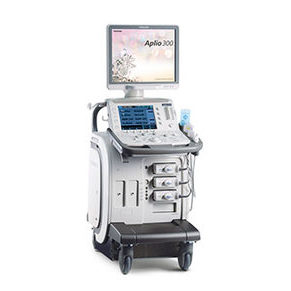
GE Logiq P9 Ultrasound Machine
The GE Logiq P9 is a mid-range ultrasound that can replace the position of Logiq P6. GE introduced Logiq P9 as a full-featured, multi-purpose diagnostic ultrasound. GE Logiq P9 is for a customer who does not want to miss the consistent image quality, comprehensive application coverage, and ease of use that enable clinicians to make timely, confident decisions.
Overview
GE Logiq P9 FAQ:
Dimensions and Weight
Height
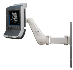
Fixed Monitor Arm (Standard)
– Maximum: 1475 mm (60.0 in)
– Minimum: 1375 mm (54.1 in)
Articulating Monitor Arm (Option)
– Maximum: 1570 mm (61.8 in)
– Minimum: 1320 mm (52.0 in)
Width
– Keyboard: 430 mm (16.9 in)
– Foot Cover: 495 mm (19.5 in)
– Monitor: 525 mm (20.7 in)
Depth
– Foot Cover: 685 mm (27.0 in)
– Rear Handle: 740 mm (29.1 in)
Weight (without peripherals)
– 60 kg/ 132 lbs
GE Logiq P9 Specifications:
Digital P-Agile Beamformer Architecture
386,469 System Processing Channels
Max. Frame Rate up to 2399 F/s
Displayed Imaging Depth: 0 – 33 cm
Minimum Depth of Field: 0 – 1 cm (Zoom, probe dependent)
Maximum Depth of Field: 0 – 33 cm (probe dependent)
270 dB of Composite Dynamic Range
Adjustable Dynamic Range
Adjustable Field Of View (FOV)
Up to 132 degrees (depending on Probe), Image Reverse: Right/Left
GE Logiq P9 Electrical power:
Voltage: 100-240 Vac
Frequency: 50/60 Hz
Power consumption maximum of 500 VA with peripherals
GE Logiq P9 Probes/Transducers:
Convex Probe
C1-5-RS [1.75-4.95Mhz]
Micro Convex Probe
8C-RS [3.6-10.0Mhz]
Endo Micro Convex Probe
E8C-RS [3.6-10.0Mhz]
Matrix Array Linear Probe
ML6-15-RS [4.3-13.0Mhz]
Linear Probe
12L-RS [3.85-11.65Mhz]
Linear Probe
9L-RS [4.5-7.8Mhz]
Linear Probe
L8-18i-RS [4.5-14.0Mhz]
Phased Array Sector Probe
3Sc-RS [1.45-4.2Mhz]
Phased Array Sector Probe
6S-RS [2.2-7.0Mhz]
Convex Volume Probe
RAB2-6-RS [1.7-4.8Mhz]
CW Split Crystal Probe
P8D [8Mhz]
GE Logiq P9 Features:
• 21.5″ widescreen LCD with high resolution
• Articulating monitor arm(option)
• 3 Active Probe Ports
• 4 Active Probe Ports(Option)
• 1 CW Pencil Probe Port
• Hard Disk Partition of 358GB for image storage without Compression
• Hard Disk Partition of 358GB for image storage without Compression
• Storage Formats
– DICOM: Compressed/uncompressed, single/multi-frame, with/without raw data
– Export JPEG, WMV(MPEG4), and AVI formats
• Advanced user interface with high resolution 10.4 inch wide LCD touch panel
• Automatic Optimization
– Auto Tissue optimization
– Auto Spectral Optimization
– Auto TGC
• CrossXBeamTM compounding
• Speckle Reduction Imaging (SRI-HD)
• Fine Angle Steer
• Coded Harmonic Imaging
• Virtual Convex
• Easy 3D
• Anatomical M-Mode
• Patient Information Database
• Image Archive on CD/DVD and Hard Drive
• Easy Backup to Media for Data Security
• TruAccess, Raw Data Processing and Analysis
• Real-time Automatic Doppler Calcs
• OB Calcs
• Fetal Trending
• Multi Gestational Calcs
• Hip Dysplasia Calcs
• Gynecological Calcs
• Vascular Calcs
• Cardiac Calcs
• Urological Calcs
• Renal Calcs
• InSiteTM ExC Capability, Remote Service
• iLinq Capability, Remote Service
• On-board electronic documentation (PDF format)
• MPEGVue
• Key Macro
• Network Storage
• Quick Save
• Quick Patient Entry
• TIC Motion Tracking
• My Page
• My Trainer
• Reset
GE Logiq P9 Peripheral Options:
• Sony Digital UP-D711 Termal Printer
• Sony Fixture Kit for Digital UP-D711 Thermal Printer
• Sony Digital UP-D25 Color Thermal Printer
• Sony Digital UP-D897 BW Thermal Printer
• Mitsubishi P93W/E Thermal Printer
• Mitsubishi P95DW Thermal Printer
• Footswitch MKF2-MED USB GP26
• USB ECG Kits (AHA/IEC)
GE Logiq P9 Supplies:
• Aquasonic ultrasound gel
• Sono ultrasound wipes
• Console Protective Cover
• Sony UPP-110HG thermal printing paper
• Sony UPC-21L color thermal printing pack
• Mitsubishi KP95HG thermal roll paper (new)
• Mitsubishi KP65HM-CE High density thermal paper
• External USB printer connection
GE Logiq P9 ports:
• HDMI Out
• Ethernet Network (RJ45)
• Wireless LAN card for wireless data transfer
• External Audio Out
• S-Video
• USB (2 x in front, 3 x in rear, 2 x monitor)
• AC Power Input
• Probe connectors
GE Logiq P9 image storage:
• Hard Disk Partition of 358GB for image storage without Compression
• Storage formats
– DICOM – Compressed /uncompressed, single/multiframe, with/without Raw Data
– Export JPEG, JPEG2000, WMV(MPEG 4) and AVI formats
• Storage Devices: USB Memory Stick
– DVD-RW Storage
– SW DVR (Option)
GE Logiq P9 Applications:
• Abdominal
• Obstetrical
• Gynecological
• Breast
• Small Parts
• Musculoskeletal
• Vascular
• Endocavitary
• Pediatric
• Neonatal
• Transcranial
• Cardiac(Adult
• Pediatric
• Stress Echo
• Intraoperative
GE Logiq P9 FAQs:
• Auto Optimization is a one-touch image optimization function that a user uses to optimize the image, based on the actual B-mode image or Pulse wave Doppler data. The function works based on preset levels (Low, Medium, and High) and allows users to pick a preference for the contrast enhancement in the resulting image. Low does the least amount of contrast enhancement, high does the most. Auto Optimization is available in single or multi image, on live, frozen or CINE images (in B-Mode only), while in zoom, and in Spectral Doppler. Auto Optimization in PW Doppler Mode optimizes the spectral data. Auto adjusts the Velocity Scale (live imaging only), baseline shift, dynamic range, and invert (if preset). Upon deactivation, the spectrum is still optimized.
• B-Flow utilizes grayscale imaging to visualize a blood flow with different gray intensities according to the reflectors’ speed and hemodynamics. B-Flow is less dependent on the user or scanning angle, while Color Doppler is heavily dependent on the scanning angle. It also provides a higher frame rate and spatial resolution than Color Flow. B-Flow may help visualize vessel-wall irregularities, kidney perfusion, liver and spleen vasculature, and bladder reflux or jets.
• SRI (Speckle Reduction Imaging) reduces speckle noise in the images, which affect the edges and fine details that limit the contrast resolution and make the diagnostic more difficult.
• CrossBeam (Spatial Compounding Imaging) obtains real time sonographic information from several different angles of insonation and combines them to produce a single image. CrossBeam helps reduce speckle artifacts, enhance mass margin delineation, and improve anatomical details.
• Coded Harmonic Imaging (CHI) utilizes Digitally Encoded Ultrasound (DEU). Harmonics enhances near field resolution for improved small parts imaging. Harmonics diminishes low frequency high amplitude noise and improves imaging for technically difficult patients. Harmonics may be especially beneficial when imaging isoechoic lesions in shallow-depth anatomy in the breast, liver, and hard-to-visualize fetal anatomy. Harmonics may improve B-Mode image quality without introducing a contrast agent.
• LogiqView (Panoramic Imaging) enables a transducer to be moved along a larger organ, stitching multiple images together to form one long image with an extremely wide field of view.
• Auto IMT automates measurement of intima-media thickness of vessels. Auto IMT helps keep track of atherosclerosis diseases from the early stage as it is developed.
• B Steer simply slants B-mode or Color Flow linear images left or right to get more information without moving the probe. This enables enhanced needle visualization in real time for needle guidance procedures. The angle steer function only applies to linear probes.
• Scan Assistant is the customable scanning assistant tool that enables a user to save scanning protocols of each lab that helps decrease key stokes from up to 60~70% of ordinary operations. With fewer user’s actions, an exam can be complete because a user does not need to move all around the console, and only requires one button to be pushed at most of the exams.
• Compare Assistant views past studies and current images together in real-time via a split-screen on the monitor.
• Strain Elastography is a non-invasive diagnostic technique that displays the relative elasticity of tissue stiffness compared to the surrounding tissue by a real-time color map that is superimposed on a conventional grayscale image. Most malignant lesions have a harder or stiffer consistency than the surrounding benign tissue. This change in stiffness can also be present in chronic or inflammatory diseases. To displace the underlying anatomical structures, Strain Elastography requires manual palpation by the user, or compression or decompression of the target produced by the patient respiration. It can be an efficient tool to improve biopsy targeting and imaging, evaluate breast and testicular lesions, and perform a quick monitoring and follow up of interventional procedures. It can also provide additional information to increase diagnostic confidence for musculoskeletal diseases such as tendinopathies, tendinosis, synovial hypertrophy, or tears.
• Auto EF is the function that automatically measures LV ejection fraction. Along with the Automated Function Imaging (AFI) feature, Auto EF maximizes time efficiency by the reproducible quantitative output that increases the physician’s diagnostic confidence.
• Tissue Velocity Imaging (TVI) is a myocardial Doppler Imaging with a color overlay on the tissue image that is available on the sector probes. Tissue color overlay can be removed to show just the 2D image, while still retaining the tissue velocity information. Tissue Velocity Imaging (TVI) is an ultrasound-based technique used for quantitative analysis of the cardiac function, and has earlier been evaluated according to myocardial velocities. The technique is the same as for Doppler echocardiography, which measures flow velocities. Tissue signals, however, have higher amplitude and lower velocities, and the signals are extracted by using different filter and gain settings.
• Battery Pack is an optional power supply. The battery maintains the system’s power when there is an AC power failure or when the power cable is unplugged. Also, this helps maintain the system’s power when it needs to be.
• Virtual Convex is available on linear and Sector probes. Virtual Convex provides a larger field of view in the far-field. Virtual Convex is always active with sector probes.